The first direct comparison of brain imaging techniques for diagnosing Alzheimer’s disease in real world patients has found that positron emission tomography (PET) is superior to single photon emission computerised tomography (SPECT).
Clinicians at the Austin Hospital, Melbourne found that cerebral blood flow 18F-FDG PET had markedly superior accuracy compared to SPECT in the assessment of people with dementia or mild cognitive impairment referred for diagnostic work-up by memory disorders specialists.
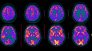 In a head-to-head comparison in 126 patients who underwent both imaging procedures they found that FDG PET was superior on almost all assessed performance measures including accuracy, sensitivity and reviewer confidence for diagnosing Alzheimer’s disease as verified by β-amyloid PET.
On the primary outcome of accuracy, FDG PET showed an overall AUROC of 0.71 compared to 0.61 for SPECT when images were reviewed by five nuclear medicine clinicians.
The investigators, led by Professor Christopher Rowe said this difference in accuracy was both clinically and statistically significant and was observed across all five reviewers irrespective of clinical experience.
FDG PET showed markedly superior sensitivity in identifying patients with Alzheimer’s disease (76% vs. 43%), whilst specificity was 74% vs 83%.