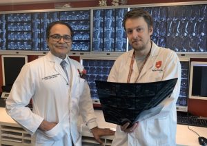
Dr Ashish Diwan and Dr Kyle Sheldrick. Picture: UNSW
A novel MRI technique developed by NSW clinicians provides better assessment of intervertebral disc degeneration, which they say could improve the diagnosis of back pain.
Dr Kyle Sheldrick, an orthopaedic spinal researcher from UNSW Medicine’s St. George & Sutherland Clinical School, says the ‘decay variance’ technique can be applied in postprocessing of current MRI technology to achieve more accurate and rapid representations of disc degeneration compared to current imaging processing techniques.
In animal studies the researchers showed that when using multi‐echo T2 MRI sequence, decay variance postprocessing had excellent correlation with degenerative changes as quantified by histological grading in a rabbit lumbar IVD degeneration model.
Decay Variance was able to separate degenerate from nondegenerate IVDs more reliably than quantitative T2 relaxometry or T2 signal intensity techniques, the .
Overall, quantitatively assess disc degeneration with an accuracy rate of 97% compared to 70% with traditional MRI methods.
Dr Sheldrick said the results were promising and could be available for human use within three years to allow radiologists to more accurately pinpoint the cause and location of a patient’s back pain.
“Current tests for disc degeneration don’t work very well. Patients with discs that look healthy on MRI often have severe back pain, and patients with discs that look very degenerate on T2 MRI often have no back pain, so better technology is needed.”
Dr Sheldrick said disc degeneration was a complex process, with a combination of lots of changes happening at a microscopic level, that are not represented clearly with current MRI techniques
“Some of the changes, like dehydration, make the individual protons line up faster while other changes, like loss of some long chains of sugars, should make them line up slower, so a really simple measure like the speed with which they line up on average won’t capture the process very well because a lot of the changes cancel each other out.”
Senior author Dr Ashish Diwan, an orthopaedic surgeon at St. George Hospital, explained that current signal decay techniques are based on how quickly or slowly individual atoms line up with a magnetic field after a strong burst of radio waves.
“Instead of trying to figure out how fast or slow this signal decay is, our technique measures whether the atoms in a sample are lining up at the same speed as each other, or a range of different speeds – hence decay variance,” he said
Dr Sheldrick said decay variance could be used on virtually all current scanners and the team will be starting a new trial inn human subjects with a Medicare licensed scanner in Sydney later in 2019.
He said it was important to improve the performance of MRI technology in the diagnosis of spinal disc degeneration because “at the moment our ability to tell where back pain is coming from is rubbish”.
“This holds back research, and our progress in treating back pain has been minimal, with 95% of people with back pain having no known cause.”
“Trying to find treatments for chest pain wouldn’t make any progress either if heart attacks, pleurisy, pericarditis, pneumonia, rib fractures, lung cancer and pneumothorax were all grouped together as ‘non-specific chest pain’. I don’t believe we will make serious progress with finding new treatments for back pain until we know what’s causing it in each person.”
Dr Sheldrick says the inability of doctors to determine the cause of back pain leads to a “one size fits all approach to medicine, in which 95% of people get the same treatment”.
“Even without finding new treatments, being able to classify people based on the cause of their back pain opens up new avenues for personalised medicine.”
“Some people might be better candidates for one exercise regimen, others might be best served by a different exercise regime, while a third group might be best served by surgery.”
The study is published in JOR Spine,