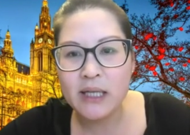
Dr Tracy Leong
The evidence of lung cancer screening benefit is now so strong that it will inevitably come to Australia, the only question being how it is implemented, according to local experts.
Presentations at the recent ERS 2020 virtual congress showed that the discussion around lung cancer screening has moved on from the major efficacy trials such as NLST and NELSON to focus on the issues of implementation and cost, says Dr Tracy Leong, Director of Bronchoscopy, Department of Respiratory and Sleep Medicine, Austin Health.
Speaking on a limbic webinar about the highlights from ERS, she said that with meta-analysis now showing a 20% reduction in lung cancer mortality with screening, many of the sessions at ERS were addressing issues such as eligibility for screening, false positives and practical questions such as whether there are enough radiologists and respiratory physician to manage the many nodules found on imaging.
Dr Leong said it was encouraging to see progress in areas such as the use of risk tools for eligibility rather than just relying on age and smoking history. And advances in CT technology mean that radiation doses have been reduced to the equivalent of three chest x-rays she noted.
Australia now awaits the results of the ILST study which has local sites, in about two year’s time, and also the outcome of a Cancer Australia inquiry into implementation of cancer screening ordered by the Minister for Health.
“So I think that screening in Australia is coming, it’s just a matter of deciding what’s the best form to put that in,” she said.
In the meantime, it is important to embrace minimally invasive and precise ways to sample the nodules that will be found on lung cancer screening, particularly as studies show more than a half will be in the peripheral lung.