Australian-developed imaging software allows easier, faster and more consistent assessment of musculoskeletal tissues and their components for conditions such as osteosarcopenia, according to Victorian specialists.
The semiautomatic volume-calculation and -rendering software Tissue Compass provides a more consistent tool to quantify musculoskeletal tissues and their components from routine CT and MRI scans, according to researchers at the Australian Institute for Musculoskeletal Science (AIMSS) at the University of Melbourne.
 Led by Dr Ebrahim Bani Hassan, a musculoskeletal pathophysiologist, and Mahdi Imani a biomedical engineer, the group has validated the system for bone and muscle analysis using 100 human CT scans.
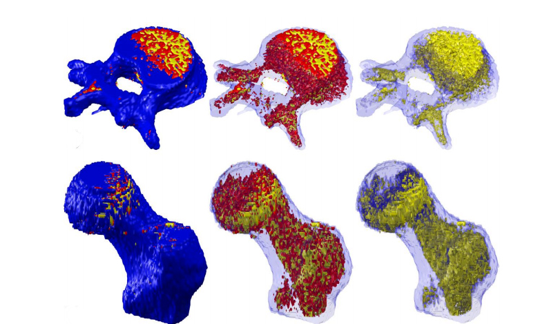
Their analysis assessed bone, hematopoietic bone marrow and marrow adipose tissue of hip and L1 spine (vertebral body) as well as muscle and intramuscular fat (iMAT) of psoas muscle in L4 spine CT scans.
The group says the system showed excellent to perfect correlation and agreement with the leading image analysis software and excellent correlation in histomorphometric validation.
There was strong correlation between the measurements of bone and marrow volume by Tissue Compass vs. tissue surface areas of in animal model histological sections. There was strong to almost perfect correlation between the system and commercial software in both bone and muscle image analysis techniques.
“This tool will give clinicians the information they need to diagnose osteosarcopenia and to act now to protect their patients’ health and independence into extreme old age,” said co-investigator Professor Gustavo Duque of the University of Melbourne.
“Fat infiltration in bone and muscle is the clearest indicator we have of osteosarcopenia and can be identified much earlier and accurately than changes to bone and muscle mass, which are used by doctors at the moment to make a diagnosis,” he said.
“These local fat levels in muscle and bone herald a range of conditions that are associated with osteosarcopenia and mostly respond very well to early treatment, such as kidney disease, vitamin D deficiency, and in men, testosterone loss. They’re also associated with fractures and falls.”
The researchers noted that clinicians have traditionally used a combination of imaging (MRIs, CT scans and densitometries) to assess muscle and bone mass, as well as grip and gait tests.
However, they said awareness of sarcopenia was low, partly due to scant guidance on what constitutes normal versus pathological mass and function loss, and how to treat it at different stages.
“There is plenty we can do through diet, exercise and medication to build up bone and muscle strength, years before people have their first fracture or fall – a moment when things can very abruptly reach a point of no return,” Professor Duque said.
“The problem we currently face is delayed diagnosis. Tissue Compass will speed up the process, allowing patients to get the right treatment for them, sooner.”
The team is now testing the system in more than 3000 participants, in collaboration with six US universities. The analysis was sponsored by AIMSS.