Some of the first images of coronavirus affected lungs have been released from post-mortem examination of patients in China, showing high levels of viscous secretions in distal airways.
The report, published on the Journal of Forensic Medicine, cover autopsies carried out by a team led by forensic pathologist Professor Liu Liang of Tongji Medical College, Huazhong University of Science and Technology, Wuhan, between February 16-24.
In his report published in the Journal of Forensic Medicine, Professor Liu says his most significant impressions of coronoavirus infection were that pulmonary fibrosis was less severe than with SARS coronavirus infection, but the symptoms of inflammation and thick exudate were more pronounced than with SARS.
He was quoted in Chinese state media as saying that coronavirus appeared to cause more deep airway and alveolar inflammatory damage than SARS.
However the damage to other organs was less apparent, though the findings were inconclusive because patients may have had co-morbid conditions.
Therefore clearing mucus in the small airways may an important aim of treatment goal. “This may affect whether tracheotomy is performed early to draw deep sputum plugs to assist respiratory support.”
A translation of the Chinese language report is provided here:
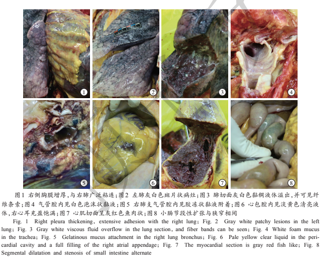
Judging from the autopsy, the lung damage of the deceased was obvious in this case. The inflammatory lesions (grey-white lesions) were weighted to the left lung, and the macroscopic view of the lungs was patchy. Grey-white lesions and dark red bleeding were visible. A spongy feeling was inherent in the lungs. A large number of sticky exudations overflowed from the alveolar surface, and fibrous strands were seen on the section. Review the CT film on the 20th day after admission, revealed multiple patchy ground glass shadows on both lungs. Air bronchograms can be seen. The left side was the most important. Fibrous cord shadows can be seen on both lower lungs.
Postmortem examination was consistent with the distribution of imaging changes, and the lesions progressed further. Considering that the ground-glass opacities seen in imaging correspond to the grey-white alveolar lesions seen with the naked eye, it suggests that COVID-19 mainly causes inflammatory reactions characterised by deep airway and alveolar damage. The local pathological changes of tissues obtained by sampling the dead body of COVID-19 deceased previously suggest that the pathological characteristics of COVID-19 are very similar to those caused by SARS and Middle East respiratory syndrome (MERS) coronavirus, but from this example the general anatomy of the system showed that the pulmonary fibrosis and consolidation were not as severe as those caused by SARS, and the exudative response was more obvious than SARS. This may be related to the short duration of this patient’s disease from diagnosis to death of only 15 days.
The pleural effusion of the deceased was not great, light yellow clear liquid, and no large amount of pleural effusion was seen, suggesting that pleural lesions were not mainly serous inflammation. The severe adhesion of the right lung to the pleura suggests that this case may be complicated by infectious pleurisy. Anatomy showed a moderate amount of light yellow clear liquid in the pericardial cavity, mild epicardial oedema, and myocardial section was grey-red, fish-like.