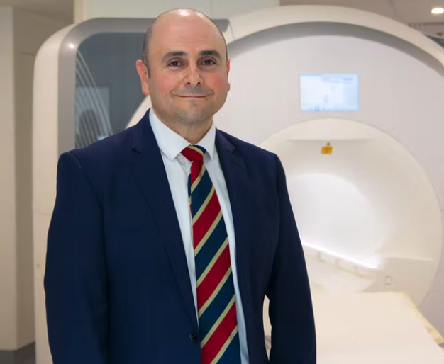
Associate Professor Andrew Jabbour
Heart transplant patients may no longer need to undergo multiple invasive biopsies to monitor rejection, according to NSW clinicians who have shown that a novel virtual MRI technique is as safe and effective in reducing post-transplant complications and hospital admissions.
Scientists at the Victor Chang Cardiac Research Institute and St Vincent’s Hospital, Sydney have published results from a pilot study showing that cardiovascular magnetic resonance (CMR)–based myocardial tissue characterisation is as effective and safe as the gold standard method of endomyocardial biopsy (EMB) for surveillance of acute cardiac allograft rejection (ACAR)-induced myocarditis in the first year after heart transplantation.
They say the non-invasive technique – which works by analysing heart oedema levels – may offer a more favourable alternative to the current requirement for 12 or more post-transplant endomyocardial biopsy procedures that are used to identify possible rejection and the need for immunosuppressive therapy.
As well as being uncomfortable, the biopsy procedure comes with a risk of rare but serious complications and both sampling error and significant interreporter variability, potentially leading to misdiagnosis and inappropriate treatment.
“It’s essential that we can monitor these patients closely and with a high degree of accuracy; now we have a new tool that can do that without the need for a highly invasive procedure,” said study investigator Associate Professor Andrew Jabbour of the Victor Chang Cardiac Research Institute.
“This new virtual biopsy takes less time, is non-invasive, more cost-effective, uses no radiation or contrast agents, and most importantly patients much prefer it,” said Associate Professor Jabbour, who is also a Consultant Cardiologist at St Vincent’s Hospital, Sydney.
In a preliminary evaluation study, 40 heart transplant patients from St Vincent’s Hospital, Sydney were randomised to receiving either a traditional biopsy or the new MRI technique. Overall, there were 401 CMR studies and 354 EMB procedures for comparison.
The results published in Circulation showed that CMR-based multiparametric assessment was highly reproducible and reliable at detecting acute cardiac allograft rejection (AUC, 0.92; sensitivity, 93%; specificity, 92%; negative predictive value, 99%) with greater specificity and negative predictive value compared to EMB-based surveillance.
High-grade rejection occurred in similar numbers of patients in each randomised group (CMR, n=7; EMB, n=8). And despite similarities in immunosuppression requirements, kidney function, and mortality between groups, the rates of hospitalisation (45% vs 90%; odds ratio, 0.091; P=0.006) and infection (35% vs 70%; OR 0.192; P=0,019) were lower in the CMR group.
Only 6% of patients randomised to the CMR arm needed a biopsy for clarification, representing a 94% reduction in the requirement for EMB-based surveillance.
The study investigators said these secondary findings are earmarked to be reconfirmed in planned larger multi-centre studies.
“The technique is now frequently used at St Vincent’s Hospital in Sydney, and I anticipate that more clinics across the world will adopt this novel technology,” commented lead investigator Dr Chris Anthony, a cardiologist at the Heart and Lung Transplant Unit, St. Vincent’s Hospital, Sydney.