New colour MRI imaging technology is allowing researchers to explore sex differences in myocardial physiology that could explain why women present with different symptoms for conditions such as acute MI.
MRI quantitative tissue mapping suggests that women have a greater degree of myocardial capillarisation than men, according to Professor Martin Ugander, the newly appointed professor of cardiac imaging at the University of Sydney.
He told the Women and Heart Disease Forum 2019 that the new MRI techniques showed that women had consistently higher levels of myocardial perfusion, blood volume and extracellular space than men, and this had implications for differential approaches to symptoms, diagnosis and treatment of heart disease in men and women.
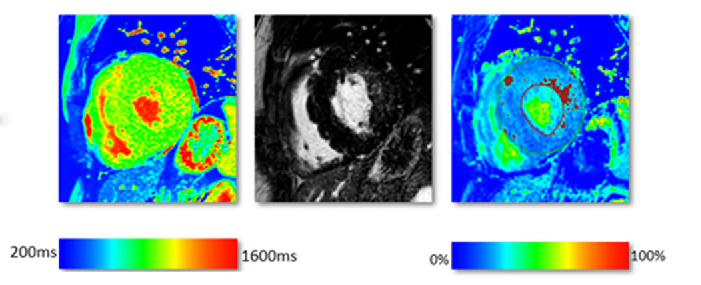
Unlike the traditional black and white MRI, the new colour MRI imaging technology now being introduced for the first time in Australia allowed objective measure of metrics such as extracellular space, myocardial perfusion and calculation of perfusion reserve, he said.
This quantitative mapping capability was also proving valuable in differentiating between different kinds of myocardial fibrosis, said Professor Ugander. MRI could tell the difference between the focal high severity scarring from myocardial ischaemia and the more diffuse fibrosis that occurs secondary to conditions such as myositis, and which may be amenable to anti-fibrotic drug therapy.
 “We now have this tool to be able to quantitatively and visually appreciate [the myocardium] and say this is microvascular disease,” he said.
Until now this has been done invasively by angiography via cath labs but MRI offered a non-invasive alternative with detailed mapping ability, he added.
“This is something we will hopefully be rolling out for broader use in the MRI community– we already have it at over 40 centres worldwide and are now installing the technology I have brought from Sweden at the North Shore Private Hospital in Sydney.”
Professor Ugander said early research using MRI quantitative mapping had shown differences in myocardial tissue between healthy male and female subjects. Women in general had 5-15% higher measures of perfusion at rest and during an adenosine stress test, and similarly higher measures of extracellular volume and myocardial blood volume.
These findings suggested that women have a higher capillary density than males and a study was now underway with the Sydney Heart Bank to further investigate these differences, he said.
“What are the implications for the symptoms that women develop, the incidence of disease and the potential for therapy? Is the higher myocardial capillary density somehow contributing to the increase in prevalence of microvascular disease in women?
“Is it contributing to differences in symptoms in women, that we’re not seeing as much chest pain but more nausea, dizziness and heart failure symptoms of dyspnoea? I don’t know, but now we have the tools using MRI to interrogate this and we see an exciting new future developing.”